
What is a brain AVM?
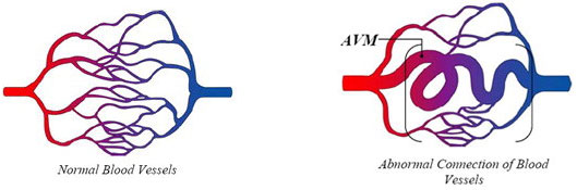
Normally, arteries carry blood containing oxygen from the heart to the brain, and veins carry blood with less oxygen away from the brain and back to the heart. When an arteriovenous malformation (AVM) occurs, a tangle of blood vessels in the brain or on its surface bypasses normal brain tissue and directly diverts blood from the arteries to the veins.
How common are brain AVMs?
Brain AVMs occur in less than 1 percent of the general population. It’s estimated that about one in 200–500 people may have an AVM. AVMs are more common in males than in females.
Why do brain AVMs occur?
We don’t know why AVMs occur. Brain AVMs are usually congenital, meaning someone is born with one. But they’re usually not hereditary. People probably don’t inherit an AVM from their parents, and they probably won’t pass one on to their children.
Where do brain AVMs occur?
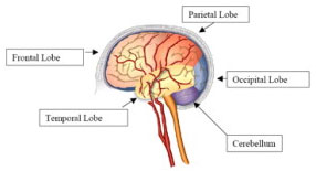
Brain AVMs can occur anywhere within the brain or on its covering. This includes the four major lobes of the front part of the brain (frontal, parietal, temporal, occipital), the back part of the brain (cerebellum), the brainstem, or the ventricles (deep spaces within the brain that produce and circulate the cerebrospinal fluid).
Do brain AVMs change or grow?
Most AVMs don’t grow or change much, although the vessels involved may dilate (widen). Some AVMs may shrink due to clots in part of the AVM. Some may enlarge to redirect blood in adjacent vessels toward an AVM.
What are the symptoms of a brain AVM?
Symptoms may vary depending on where the AVM is located:
What causes brain AVMs to bleed?
A brain AVM contains abnormal and, therefore, “weakened” blood vessels that direct blood away from normal brain tissue. These abnormal and weak blood vessels dilate over time. Eventually they may burst from the high pressure of blood flow from the arteries, causing bleeding into the brain.
What are the chances of a brain AVM bleeding?
The chance of a brain AVM bleeding is 1 percent to 3 percent per year. Over 15 years, the total chance of an AVM bleeding into the brain — causing brain damage and stroke — is 25 percent.
Does one bleed increase the chance of a second bleed?
The risk of recurrent intracranial bleeding is slightly higher for a short time after the first bleed. In two studies, the risk during the first year after initial bleeding was 6 percent and then dropped to the baseline rate. In another study, the risk of recurrence during the first year was 17.9 percent. The risk of recurrent bleeding may be even higher in the first year after the second bleed and has been reported to be 25 percent during that year. People who are between 11 to 35 years old and who have an AVM are at a slightly higher risk of bleeding.
What can happen if a brain AVM causes a bleed?
The risk of death related to each bleed is 10 percent to 15 percent. The chance of permanent brain damage is 20 percent to 30 percent. Each time blood leaks into the brain, normal brain tissue is damaged. This results in loss of normal function, which may be temporary or permanent. Some possible symptoms include arm or leg weakness/paralysis, or difficulty with speech, vision or memory. The amount of brain damage depends on how much blood has leaked from the AVM.
What functions does an AVM affect?
If an AVM bleeds, it can affect one or more normal body functions, depending on the location and extent of the brain injury. Different locations in the brain control different functions:
Are there different types of brain AVMs?
All blood vessel malformations involving the brain and its surrounding structures are commonly referred to as AVMs. But several types exist:
• Dural carotid cavernous sinus fistula. These occur behind the eye and usually cause symptoms because they divert too much blood toward the eye. Patients have eye swelling, decreased vision, redness and congestion of the eye. They often can hear a “swishing” noise.
• Transverse-Sigmoid sinus dural fistula. These occur behind the ear. Patients usually complain of hearing a continuous noise (bruit) that occurs with each heartbeat, local pain behind the ear, headaches and neck pain.
• Sagittal sinus and scalp dural fistula. These occur toward the top of the head. Patients complain of noise (bruit), headaches, and pain near the top of the head; they may have prominent blood vessels on the scalp and above the ear.
What is the best treatment for a dural fistula?
The best treatment is usually endovascular surgical blocking of the abnormal connections that have caused the fistula. This involves guiding small tubes (catheters) inside the blood vessel with X-ray guidance and blocking off the abnormal connections. Depending on the location and size, many of these can be treated and cured by these less invasive endovascular techniques.
How are AVMs diagnosed?
Most AVMs are detected with either a computed tomography (CT) brain scan or a magnetic resonance imaging (MRI) brain scan. These tests are very good at detecting brain AVMs. They also provide information about the location and size of the AVM and whether it may have bled. A doctor may also perform a cerebral angiogram. This test involves inserting a catheter (small tube) through an artery in the leg (groin). Then it’s guided into each of the vessels in the neck going to the brain, and a contrast material (dye) is injected and pictures are taken of all the blood vessels in the brain. For any type of treatment involving an AVM, an angiogram may be needed to better identify the type of AVM.
What factors influence whether an AVM should be treated?
In general, an AVM may be considered for treatment if it has bled, if it’s in an area of the brain that can be easily treated and if it’s not too large.
What is the best treatment for an AVM?
It depends on what type it is, the symptoms it may be causing and its location and size.
What different types of treatment are available?
What doctors specialize in treating brain AVMs?
Neuroradiologists specialize in the diagnosis and imaging of the head, neck, brain and spinal cord. They perform and interpret the CT, MRI, and cerebral angiograms necessary for evaluation, management and treatment. Each of these specialists has had advanced training and is highly skilled at treating complex brain vascular malformations.


