
Cerebral Venous Thrombolysis

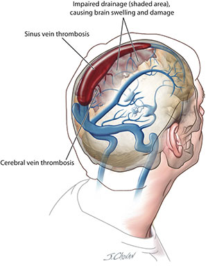
Thrombosis of cerebral veins or venous sinuses is a much less common cause of cerebral infarction than that caused by arterial disease. Clinically the diagnosis can be very difficult but modern imaging techniques allow earlier diagnosis and the possibility of early treatment. It is more likely to occur in patients with a prothrombotic tendency (for example, in pregnancy) who also have local infection (for example, sinusitis) and are either dehydrated, or have widespread malignancy. Headache and seizures are common.
Aetiology

There are many possible contributory factors and more than one may be present:

Infection is well recognised with spread from sinuses.
Trauma can result in cerebral sinus thrombosis, as can some neurosurgical procedures.
Pregnancy, the puerperium and the combined oral contraceptive pill are associated with a predisposition to venous thrombosis, including intracranial venous thrombosis.
Hypercoagulable states associated with the antiphospholipid syndrome and thrombophilia, including protein S and C deficiencies, antithrombin III deficiency, lupus anticoagulant, and the Leiden factor V mutation increase risk. Congenital varieties of thrombophilia account for around 15% of cases.
Crohn's disease and ulcerative colitis increase the risk, as does the use of steroids to treat them.
Haematological conditions include paroxysmal nocturnal haemoglobinuria, thrombotic thrombocytopenia purpura, sickle cell disease and polycythaemiarubravera. Some malignancies are associated with increased risk.
Vascular diseases include systemic lupus erythematosus (SLE), Wegener's granulomatosis and Behçet's disease. A survey from Saudi Arabia found Behçet's syndrome to be a common aetiological factor.
Risk is also increased by nephrotic syndrome, dehydration, cirrhosis and sarcoidosis.
Infection, trauma and tumour account for about 10% of cases and the importance of infection appears to be declining.
Presentation

Occlusion of the cerebral veins or dural venous sinuses may present as a stroke syndrome, subarachnoid haemorrhage or as isolated raised intracranial pressure.

Headache is a common presenting symptom.[4] It may be sudden like a blow to the head as in subarachnoid haemorrhage. There may be nausea and vomiting too. Headache can be the only feature with no neurological deficit. Headache can also be a feature that precedes stroke by a matter of weeks.
There may be seizures and even status epilepticus. About 40% of patients develop epileptic seizures at the onset or during the first days after thrombosis occurs.
Impaired level of consciousness is common but it may be normal, there may be confusion or it may progress to coma.
Possible neurological signs include hemiparesis, weakness of the lower limbs, sometimes bilaterally, aphasia, ataxia, chorea and hemianopia.
Specific cranial nerve lesions can include vestibular neuropathy, pulsatile tinnitus, unilateral deafness, diplopia, facial weakness and obscuration of vision. If the thrombosis involves the jugular vein, the IX, X, XI and XII nerves may be involved.
Papilloedema may be seen.
Investigations
The diagnosis is usually made with the aid of CT or MRI scanning. The picture shows an area of infarction that is not compatible with arterial occlusion.
Presentation

This will be determined by many factors. The neurological complications, the cause and any associated diseases need to be considered.

General measures appropriate to the rehabilitation of stroke patients. Elevation of the head at 30-40° helps to reduce intracranial pressure.
Seizures should be treated with anticonvulsants.
Specific treatment involves anticoagulation or thrombolytic treatments.

The predictors of poor outcome include coma, ICH, rapidly progressing clinical deficits, posterior fossa lesions, and involvement of the deep venous system. Approximately 30% of patients with 1 or more of these risk factors had poor outcome despite treatment with heparin.

For these patients, endovascular thrombolysis might give better results. Failure to respond to adequate systemic anticoagulation has been considered as a possible indication of endovascular thrombolysis. Evidence from small case series suggests that local thrombolytic therapy is relatively safe and effective in rapid recanalization of thrombosed sinus and reversal of neurologic deficits.4,5Local intrasinus thrombolysis dissolves the thrombus by infusion of a thrombolytic drug into the occluded sinus. The theoretic advantage of intrasinus thrombolysis for cerebral venous and sinus thrombosis is that the drug is delivered where needed.

Complications

It is important not to miss the diagnosis of intracranial venous thrombosis, as it can result in death and disability. Complications can arise from both the neurological consequences and also from the associated contributory diseases. Complications include seizures, hydrocephalus, intracranial hypertension and neurological deterioration

Prognosis
Long-term follow-up suggests a generally good prognosis but there may be residual pyramidal symptoms, epilepsy, visual field defects and depression.
Recurrence rates of 2.8% and mortality rates of 10% have been reported, despite anticoagulation treatment.
Risk factors for a poorer prognosis and increased mortality include being female, infection, hydrocephalus, intracranial haemorrhage and motor deficits.
Earlier diagnosis using angiography and MRI followed by effective anticoagulation has greatly improved the prognosis.
Submit Query
Contact form submitted! We will be in touch soon.

Submit*Required fields

Contact :

info@irsurat.com

+91 97254 01144, +91 99250 24869

Follow me on